Introduction to calcium imaging lab
🧠
Daniel Fürth
Assistant professor
SciLifeLab/Uppsala University
🔗 furthlab.xyz
@furthlab
based on material by Ashley Juavinett’s
Allen Institute Data Lesson Plans
Calcium imaging

Cre driver lines
- The Cre/lox system can also be used to produce strains in which a transgene is either inducible or expressed only in certain tissues.
Cre driver lines
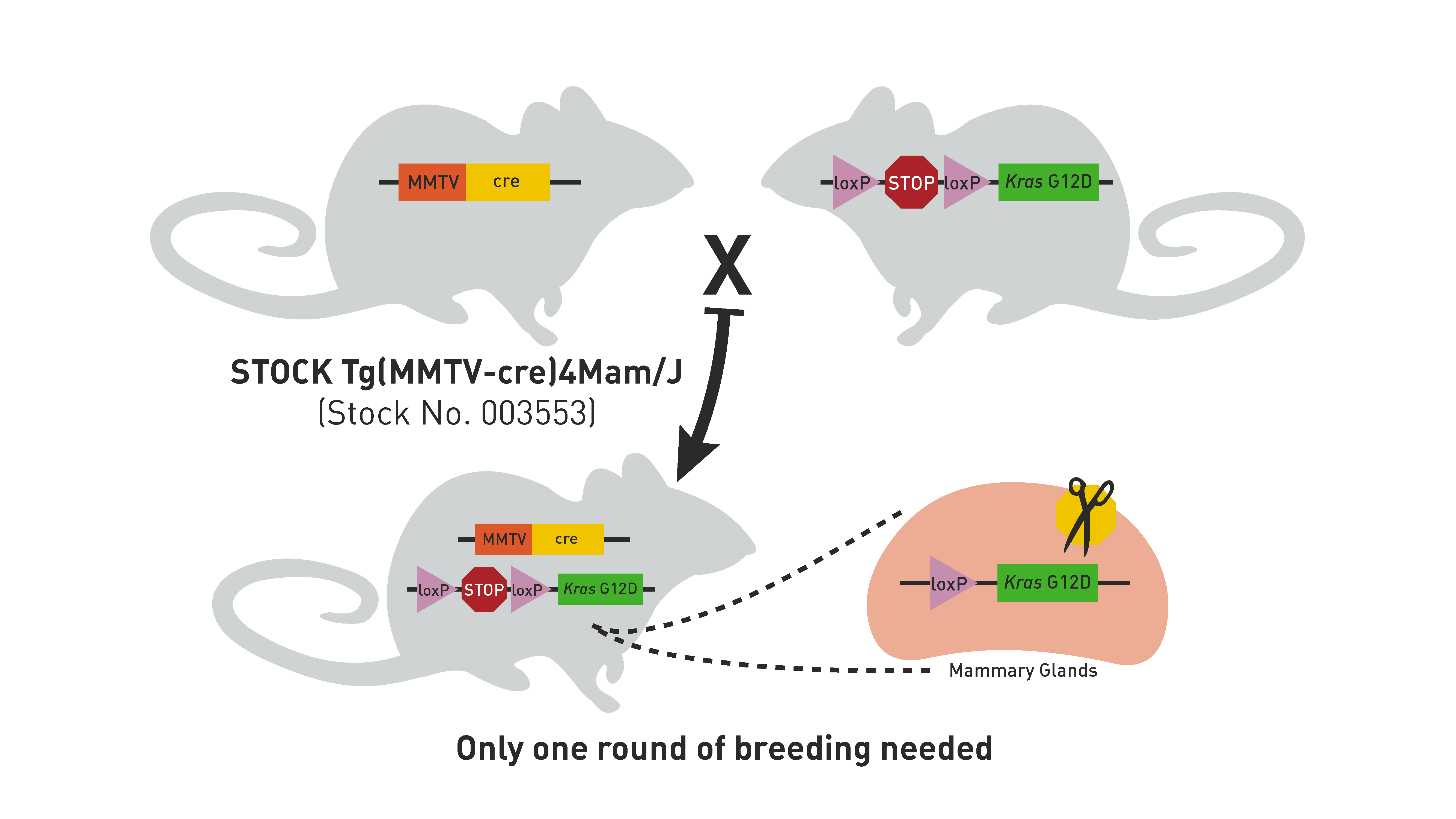
MMTV is expressed mainly in mammary glands.
Cre driver lines
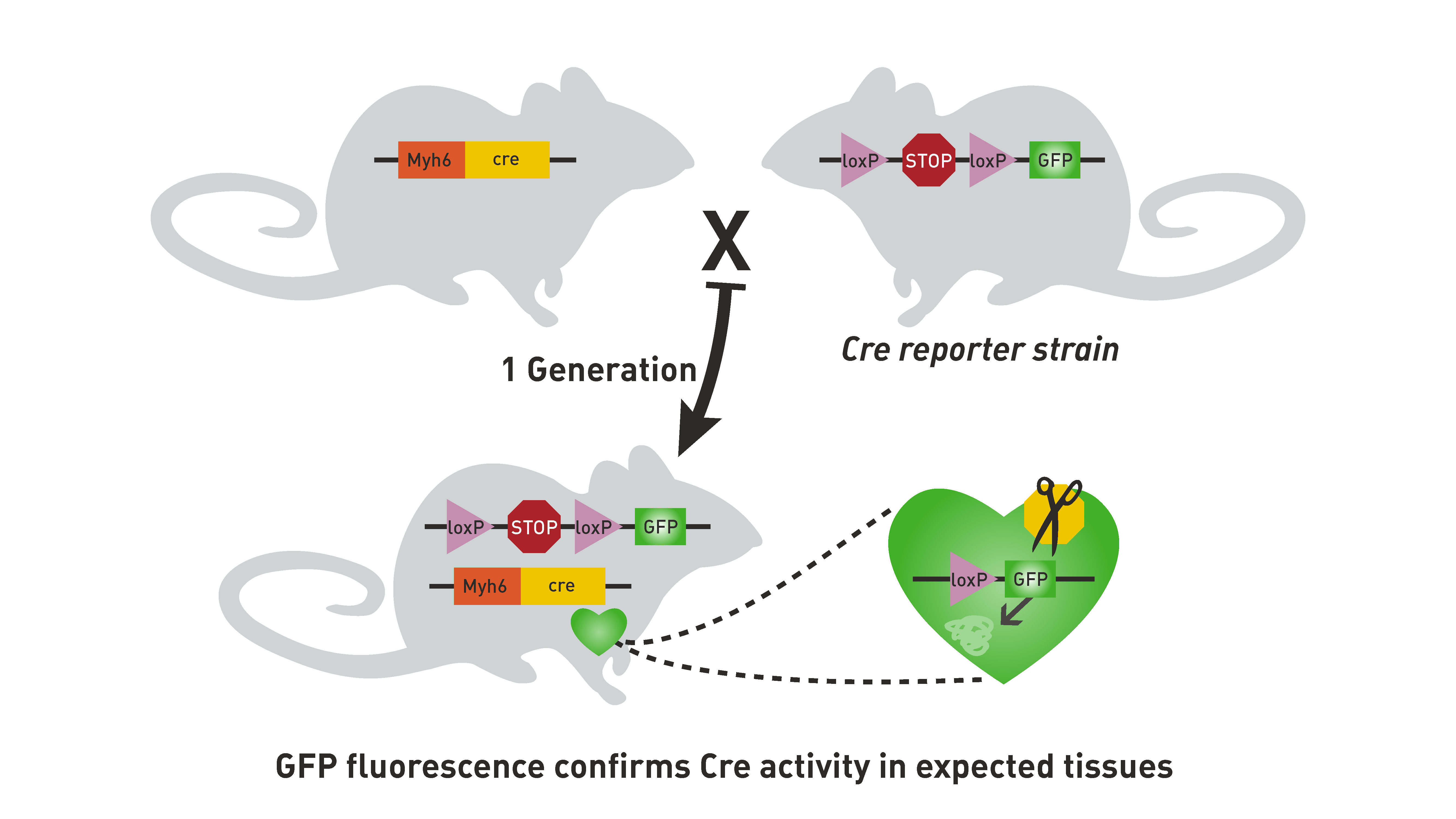
Researchers needed way to confirm that Cre recombinase was active only in certain tissues.
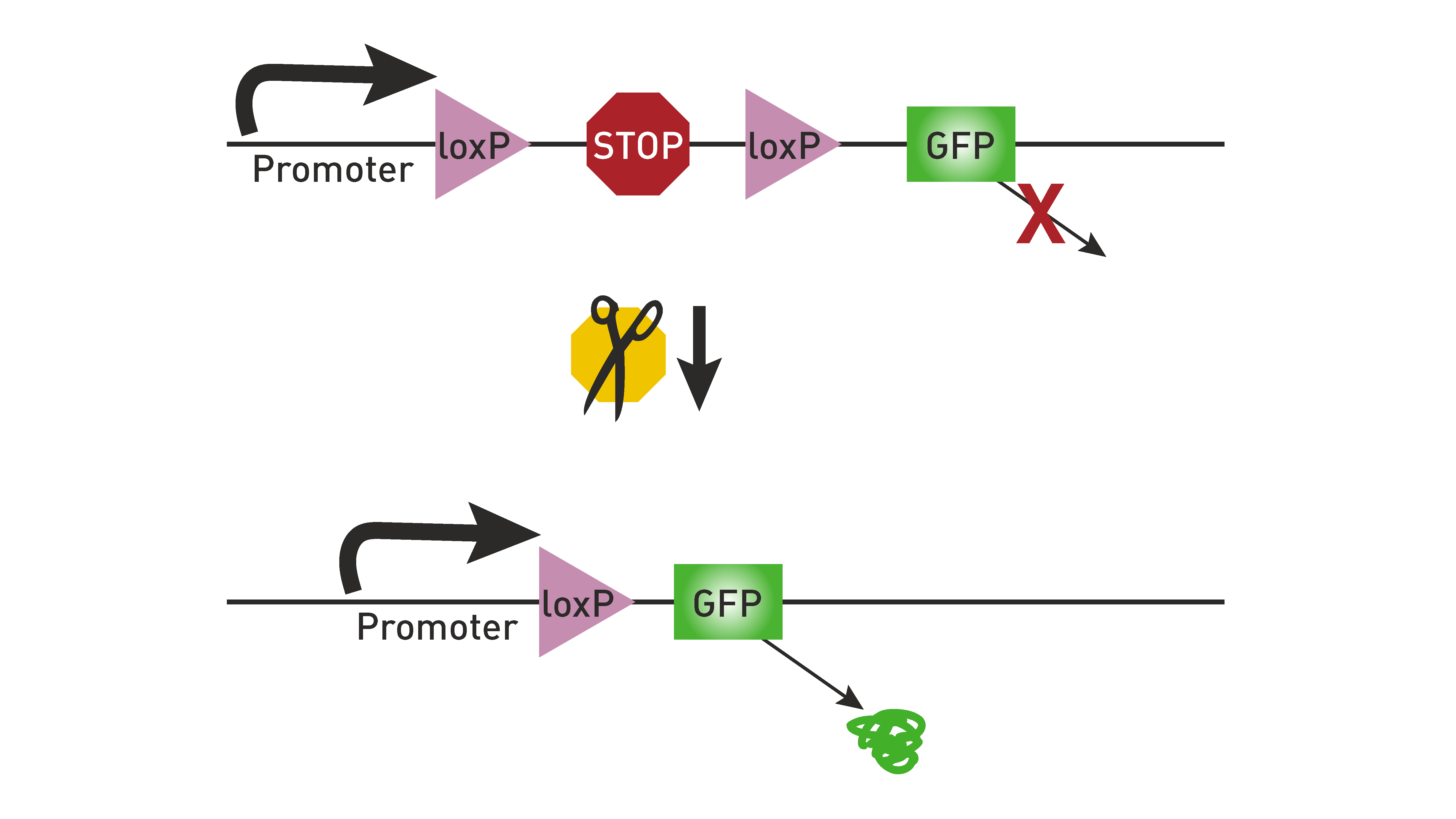
This need led to the development of Cre reporter strains.
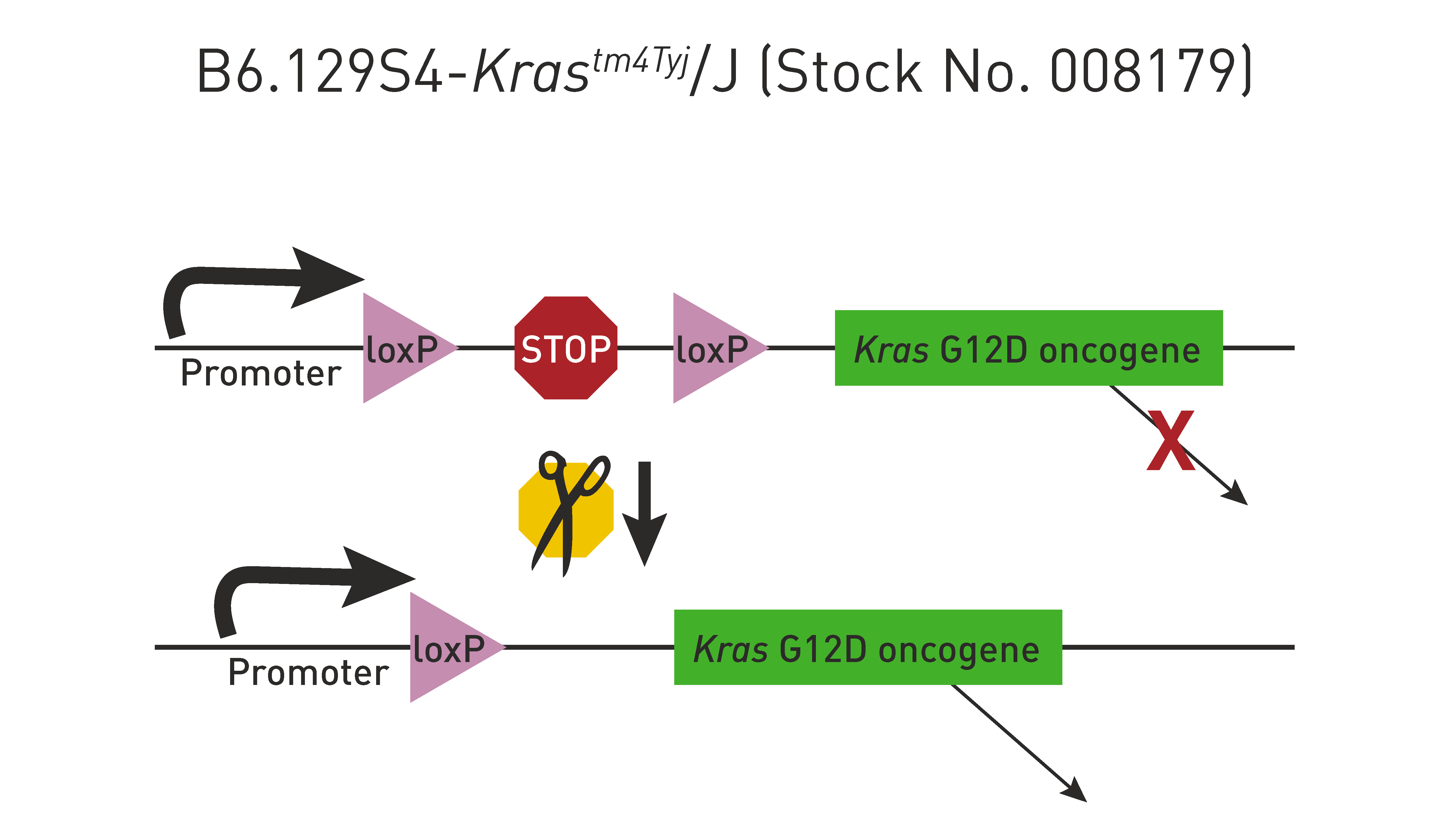
Express a visible marker, such as green fluorescent protein (GFP) or LacZ, only after Cre recombinase excises a loxP-flanked stop sequence
Cre driver lines
Recombinase: enzyme that catalyse site-specific recombination events within DNA.
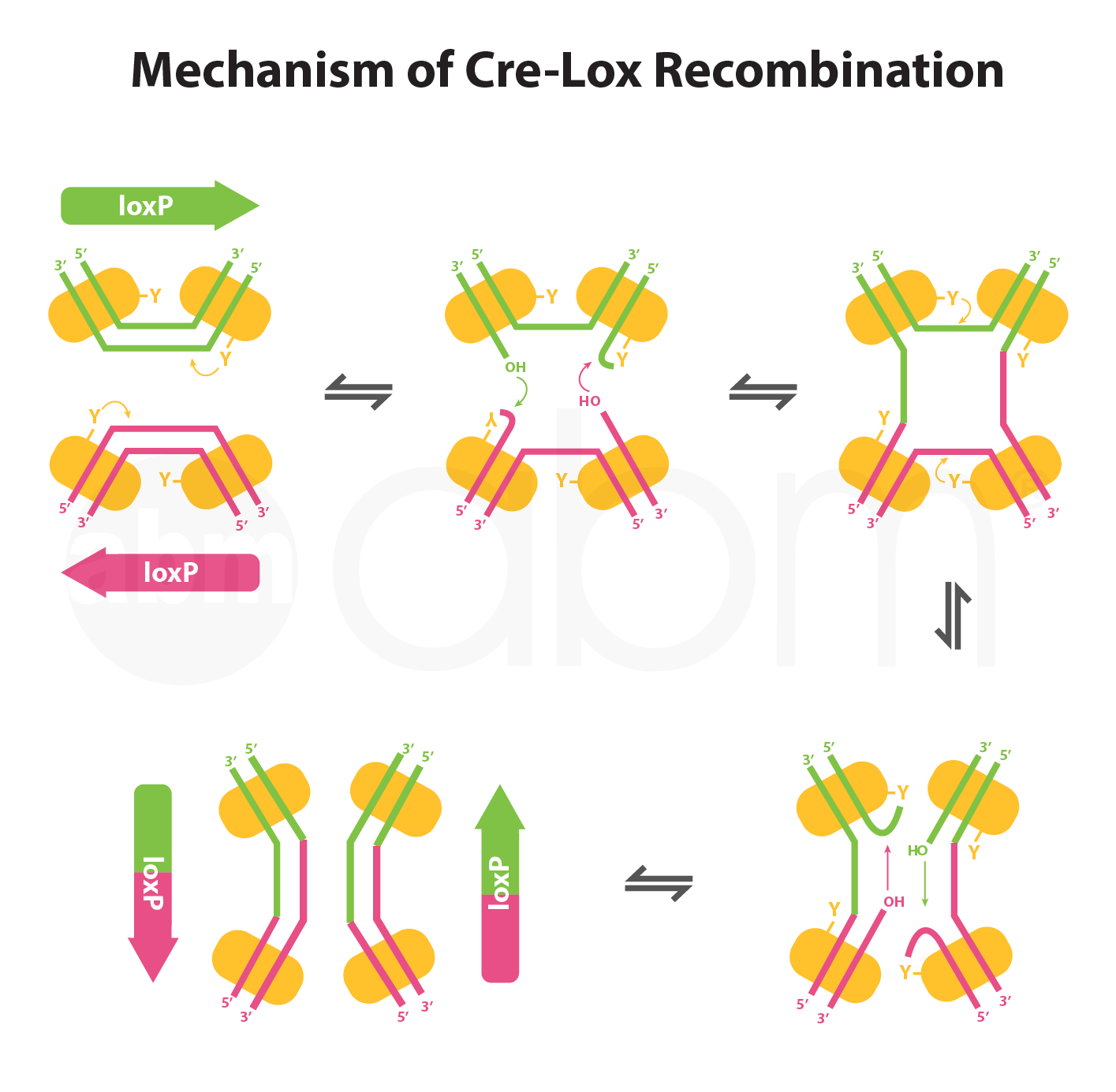
Cre proteins recognize a loxP site and bind to it, forming a dimer.
Two Cre-loxP dimers come together to form a tetramer, bringing the two loxP sites together with opposing directionality.
dsDNA cleavage occurs in the center of the loxP site and a crossover event occurs
Cre driver lines
DIO: Double-floxed gene with Inverted Orientation.
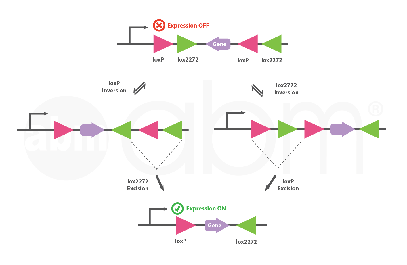
DIO vector: a Double-floxed gene with Inverted Orientation.
Inversion occurs via either loxP or lox2272, followed by the excision of two lox sites.
The DIO vector turns on gene expression only in the presence of Cre.
Cre driver lines
- Jackson Laboratory
- GENSAT
GENSAT

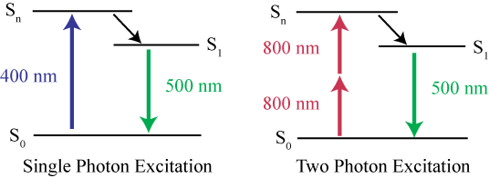
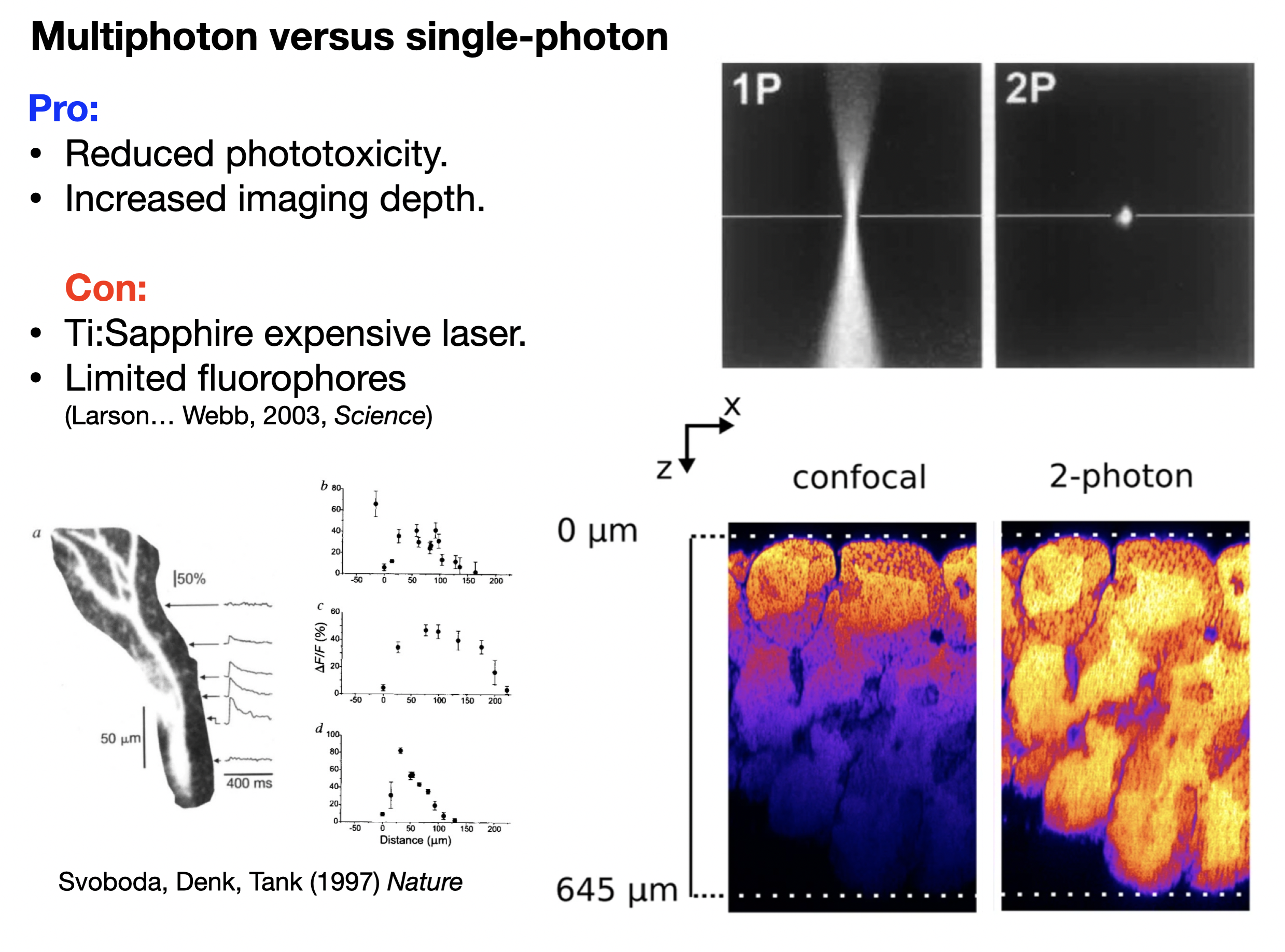
Two photons are better than one
In standard fluorescent (epifluorescence) and confocal microscopy, one photon excites the fluorophore
In two-photon microscopy, the fluorophore absorbs two photons simultaneously. With two photons - we can use half of the energy!
Half of the energy = twice the wavelength (ends up being infrared light)
Two photons are better than one
Genetically encoded calcium indicators
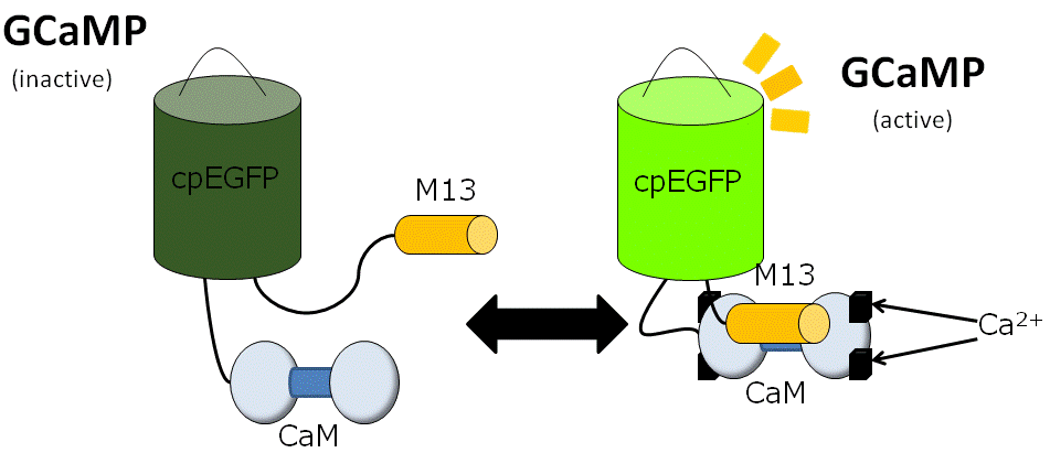
- genetically encoded calcium indicator (GECI)
- 🟢 green fluorescent protein (GFP)
- 🔵 calmodulin (CaM), a calcium-binding messenger protein
- 🟠 M13, a peptide sequence from myosin light-chain kinase.
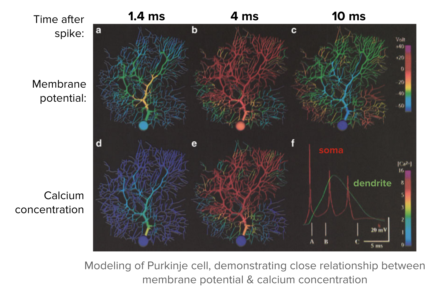
Calcium levels and membrane potential
Calcium indicators can be dyes or fluorescent proteins
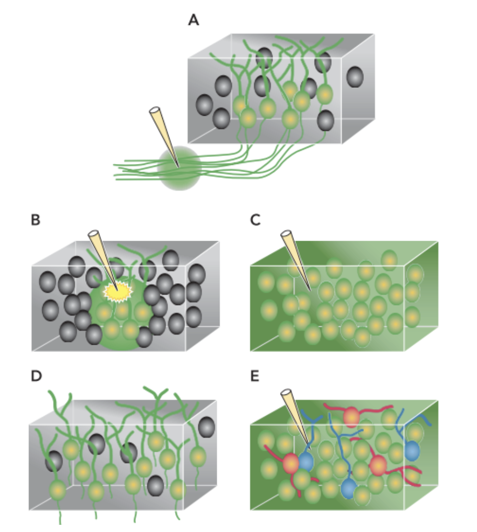
Different ways to deliver or expressed.
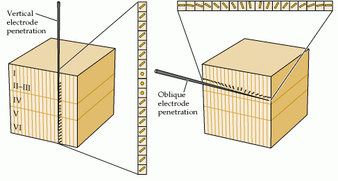
A-C different types of bulk loading (including with an electrical current in B)
D Genetically-encoded & targeted (via viruses)
E In combination with other fluorescent dyes
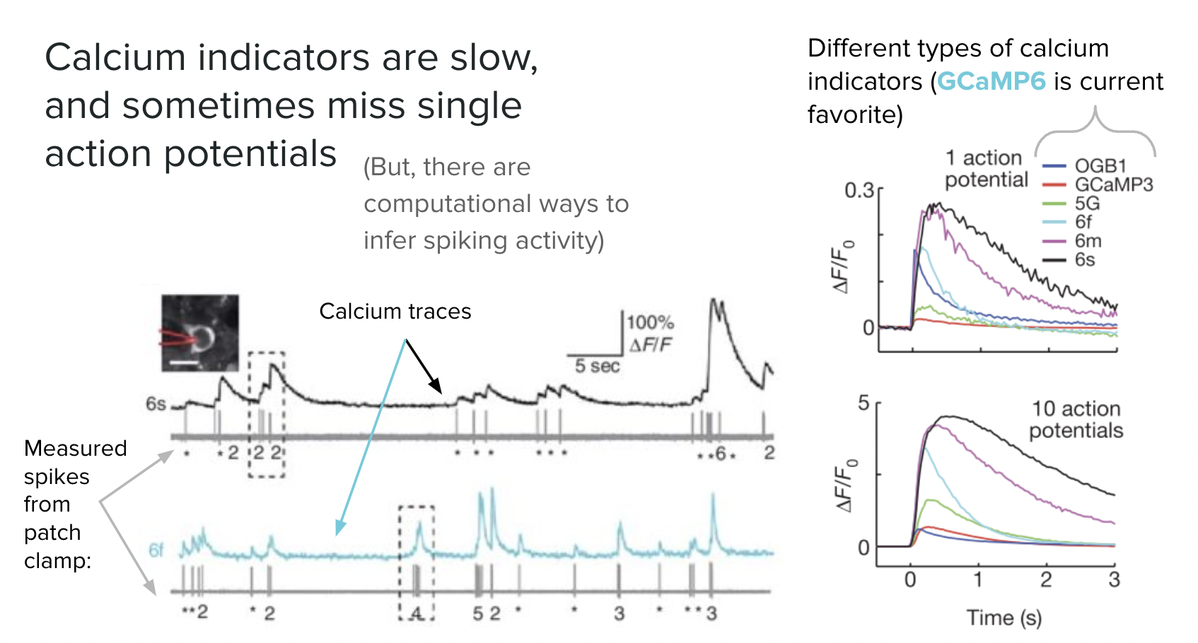
Calcium waves and spikes
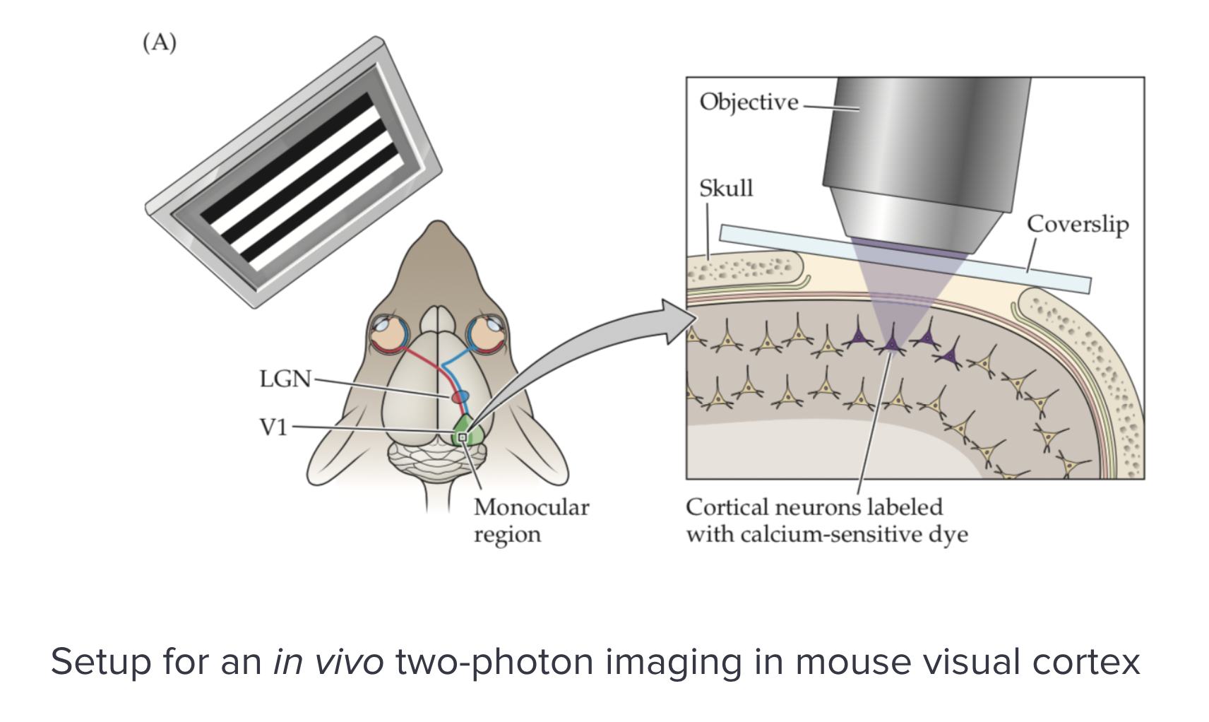
In vivo calcium imaging
In vivo calcium imaging
Direction sensitivity in vision cortex
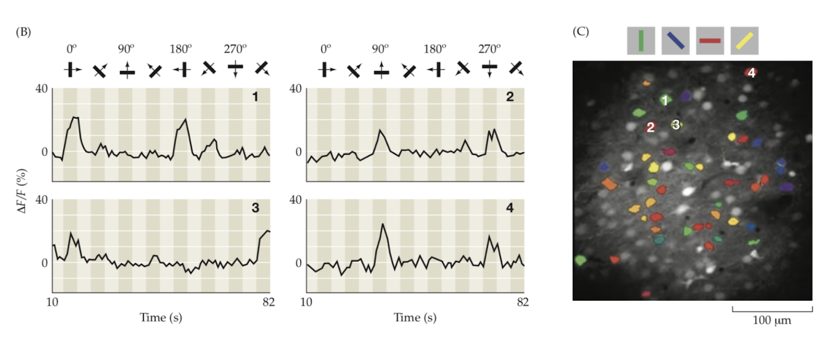
Direction sensitivity in vision cortex